
Amniotic Membrane in OB/GYN
As regenerative medicine and tissue engineering technologies continue to advance, amniotic membrane has emerged as an important biomaterial in obstetrics and gynecology. Rich in collagen, laminin, fibronectin, and various growth factors, it has demonstrated significant benefits in reducing postoperative adhesions, inflammation, and pain.
Following standardized procedures—including donor screening, tissue processing, cryogenic preservation, storage, and terminal sterilization—amniotic membrane can be prepared in sheets or other formats for use as a tissue covering and repair material in gynecologic surgery.
One of the key features of amniotic membrane is its natural three-dimensional extracellular matrix, which provides a favorable microenvironment for cell attachment, migration, and tissue remodeling. Its soft texture and excellent conformability allow it to adapt to the irregular surfaces of the uterus, vagina, and pelvic tissues, making it suitable for delicate gynecologic procedures. Its natural barrier properties help separate adjacent tissues, reduce direct tissue contact after surgery, and provide a supportive interface for tissue repair.
In intrauterine surgery, amniotic membrane may be used to cover damaged tissue following adhesiolysis. Intrauterine adhesions are commonly associated with endometrial injury, miscarriage-related procedures, infection, or other intrauterine interventions. Severe adhesions may affect menstruation, embryo implantation, and fertility. After adhesiolysis, placing an amniotic membrane over the injured uterine surface may serve as a temporary biological barrier, help maintain the healing space, and support endometrial repair and regeneration. Depending on the patient’s condition, it may be combined with an intrauterine stent, hormonal therapy, and postoperative follow-up as part of a comprehensive treatment strategy.

Common gynecologic procedures may involve vaginal defects, vaginal stenosis, congenital vaginal abnormalities, and soft-tissue reconstruction following tumor removal or trauma. AmnioLink® features flexibility, conformability, and resistance to shrinkage after hydration, allowing it to be shaped and applied according to the size of the defect. Its extracellular matrix supports the migration of surrounding cells into the repair area, promoting epithelial coverage and tissue integration while providing greater flexibility during reconstructive procedures.
In pelvic and reproductive-organ surgery, amniotic membrane may also serve as an anti-adhesion barrier in procedures involving myomectomy, uterine repair, fallopian-tube surgery, ovarian surgery, and other pelvic operations. Postoperative adhesions may lead to chronic pelvic pain, restricted organ mobility, or impaired fertility. Covering the surgical site with amniotic membrane may reduce direct friction and adhesion between injured surfaces and adjacent tissues while supporting postoperative tissue repair.
Amniotic membrane also has potential clinical value in the management of perineal wounds, vaginal mucosal injuries, and postpartum soft-tissue repair. Its thin and flexible structure conforms closely to the wound, helping protect the repair site and maintain a supportive tissue environment.
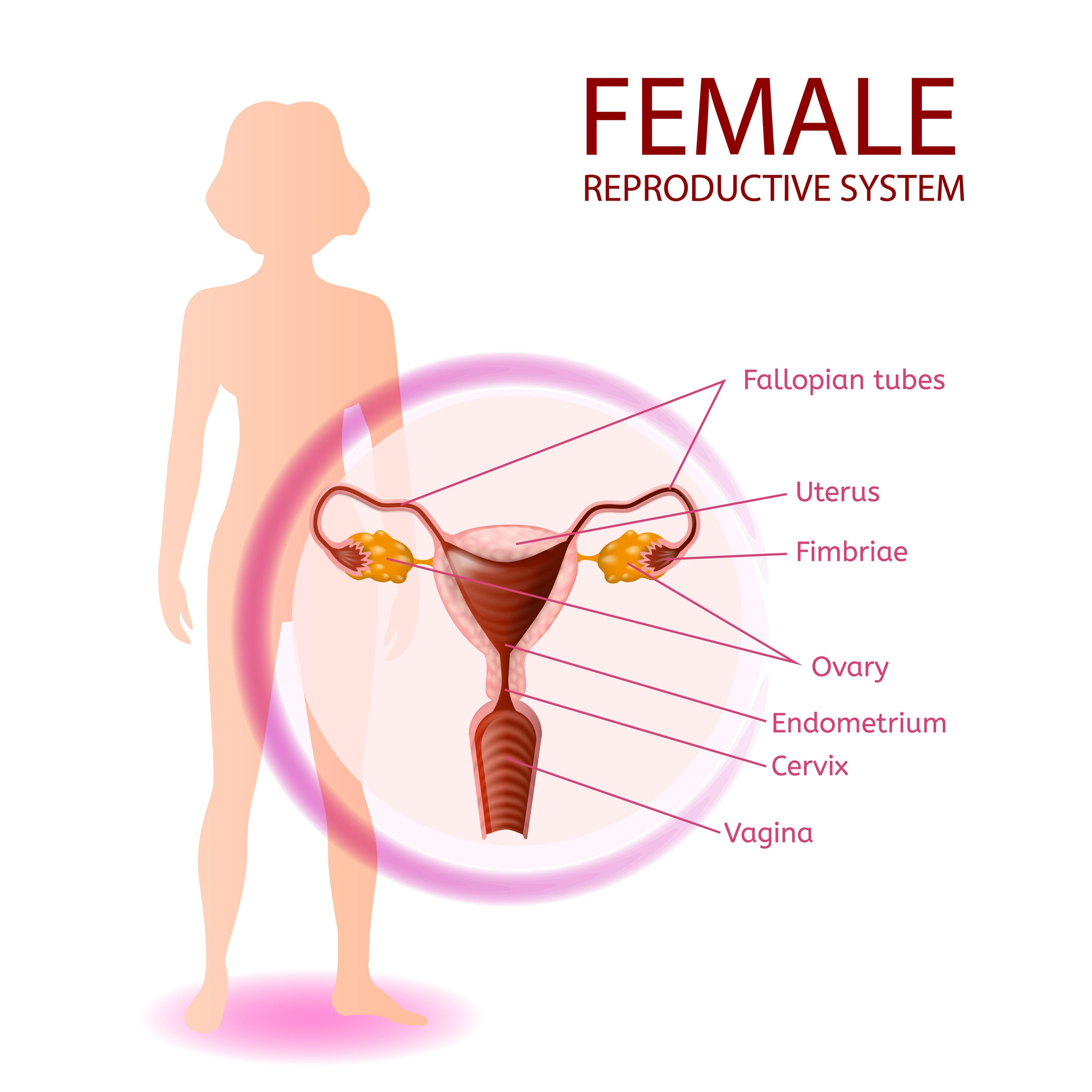
Overall, amniotic membrane combines a natural extracellular matrix with biological barrier properties, flexibility, conformability, and ease of shaping, making it suitable for a wide range of intrauterine, vaginal, perineal, and pelvic procedures. With standardized processing, rigorous quality control, and appropriate clinical application, amniotic membrane can serve as an important biomaterial for tissue repair and reconstruction in obstetrics and gynecology.

