
Amniotic Tissue in Orthopedics
As regenerative medicine and biomaterial technologies continue to advance, amniotic tissue has become a biomaterial of growing interest in orthopedics, sports medicine, and rehabilitation medicine. Located in the innermost layer of the placenta, the amnion consists of an epithelial layer, basement membrane, and extracellular matrix (ECM). It naturally contains collagen, laminin, fibronectin, and various bioactive factors. Following tissue processing, freeze-drying, and sterilization, amniotic tissue can be prepared as sheets, membranes, or micronized matrices for surgical coverage, tissue wrapping, repair-interface protection, and minimally invasive applications.
The principal value of amniotic tissue lies in its natural three-dimensional ECM structure, which provides a microenvironment that supports cell attachment, migration, and tissue remodeling. Its biological barrier properties may help separate the repair site from surrounding tissues while supporting the modulation of local inflammation, fibrosis, angiogenesis, and epithelial repair. These characteristics have led to its investigation in the management of tendon, ligament, joint, and soft-tissue injuries.

In rotator cuff repair, an amniotic membrane may be placed over the repaired tendon or at the tendon–bone interface to provide an ECM scaffold and protect the repair site. In large, degenerative, or recurrent tendon tears, it may serve as an adjunctive covering material to support the local healing environment, collagen organization, and tissue integration.
In Achilles tendon and other lower-extremity tendon repairs, an amniotic membrane may be wrapped around the sutured area to form a soft, conformable biological barrier. This may help reduce direct friction and adhesion between the repaired tendon and surrounding tissues. Similar applications have been explored in repairs involving the patellar, quadriceps, posterior tibial, and peroneal tendons to help preserve the tendon-gliding space and support postoperative functional recovery.
Because the flexor and extensor tendons of the hand are located close to tendon sheaths, nerves, and blood vessels, postoperative adhesions may restrict finger movement. An amniotic membrane may be wrapped around the repaired tendon or positioned between the tendon and tendon sheath to create a tissue-separation layer. It may therefore serve as an adjunct in hand-tendon repair and adhesion-release procedures.
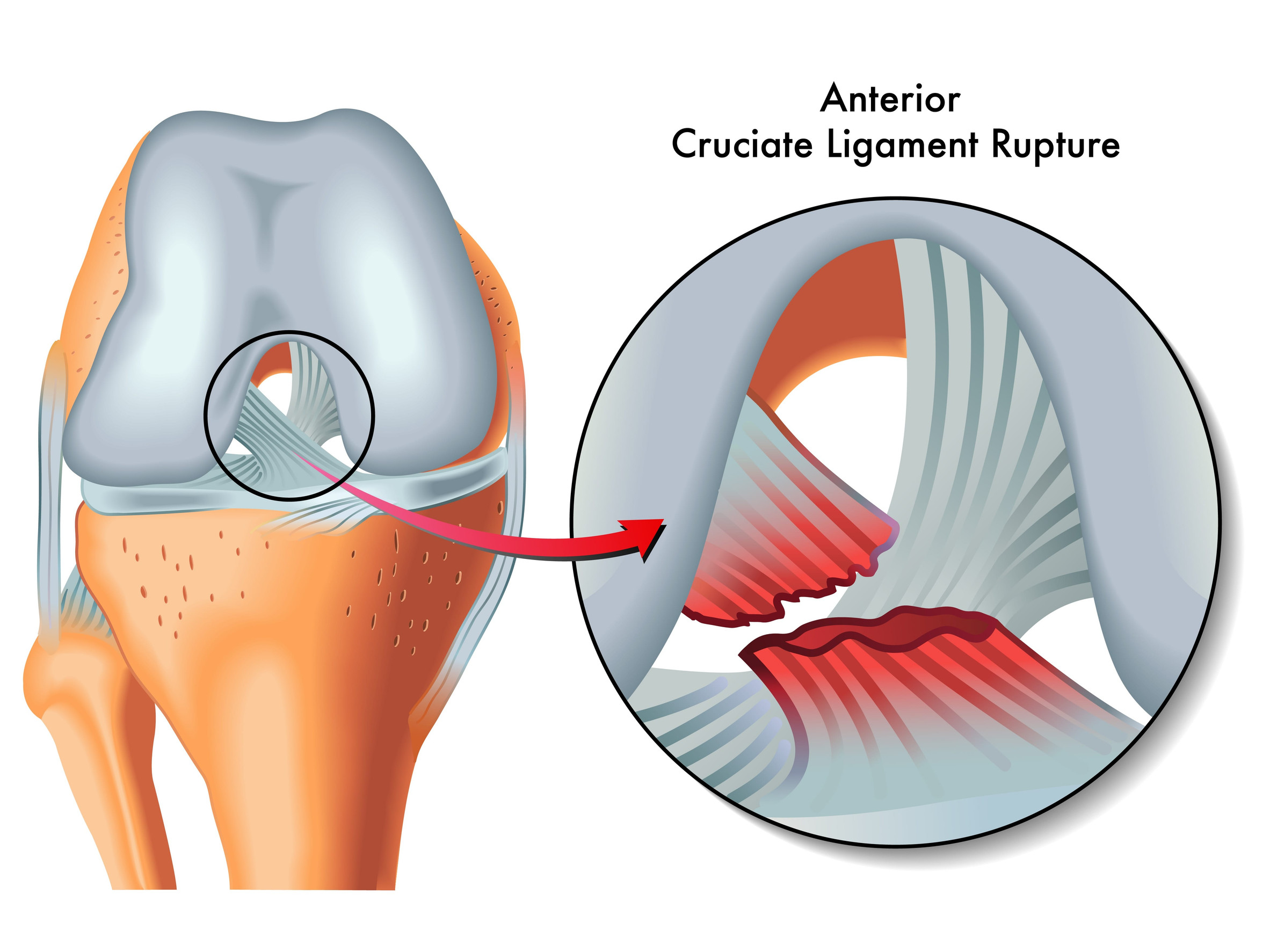
In anterior cruciate ligament, posterior cruciate ligament, and collateral ligament repair or reconstruction, an amniotic membrane may be wrapped around the graft or placed over the repair site. Its ECM structure provides an environment that may support cell migration and tissue remodeling. When applied at the interface between a ligament graft and bone tunnel, it may also support local tissue integration and protect the surrounding soft-tissue environment.
For knee osteoarthritis, cartilage degeneration, and chronic periarticular pain, micronized amniotic tissue may be considered, subject to physician assessment and applicable regulations, as a tissue matrix for the joint or surrounding soft tissues. Research has focused on supporting the joint environment, modulating local inflammation, and managing pain and physical function. Such applications may be incorporated into an integrated care plan that includes rehabilitation, therapeutic exercise, and other conservative treatments.
Amniotic membrane has also been explored in fracture fixation, joint replacement, orthopedic wound repair, and revision surgery. Its soft, conformable structure and resistance to shrinkage after hydration allow it to cover irregular surfaces and be positioned around fixation plates, screws, suture sites, nerves, and tendons. As a biological barrier, it may help maintain a favorable tissue microenvironment and reduce the effects of postoperative scarring and adhesions on tissue mobility.
Overall, amniotic tissue is not intended to replace bone, cartilage, tendon, or ligament. Instead, it serves as an adjunctive biomaterial within orthopedic repair strategies. Through its natural ECM structure, biological barrier properties, and tissue conformability, it may support tendon and ligament repair, joint care, nerve protection, and postoperative adhesion management, providing additional options for orthopedic surgery and minimally invasive treatment.

