
Clinical Use of Amniotic Tissue in Otolaryngology
Otolaryngology involves essential physiological functions such as hearing, breathing, swallowing, and voice production. Many surgical sites in this specialty are covered by mucosa or thin tissue layers. When tissue defects occur in the nasal cavity, paranasal sinuses, tympanic membrane, oropharynx, larynx, or trachea because of disease, trauma, or surgery, promoting re-epithelialization, reducing adhesions, and preserving function become important clinical considerations.
With its natural extracellular matrix (ECM), retained growth factors, favorable biocompatibility, flexibility, conformability, and absorbability, amniotic tissue may serve as a wound covering, tissue-separation barrier, and repair scaffold. These properties make it a potential biomaterial for supporting both structural and regenerative requirements in delicate otolaryngological procedures.
A key characteristic of amniotic tissue is its natural three-dimensional ECM, which provides a microenvironment that may support cell attachment, migration, and tissue remodeling. Its flexible structure allows it to conform to irregular anatomical surfaces within the nasal cavity, sinuses, middle ear, pharynx, and larynx. It may also be trimmed and secured according to the size and location of the surgical defect. As a biological barrier, it can separate adjacent injured surfaces and may help reduce direct wound contact and excessive fibrous adhesion.
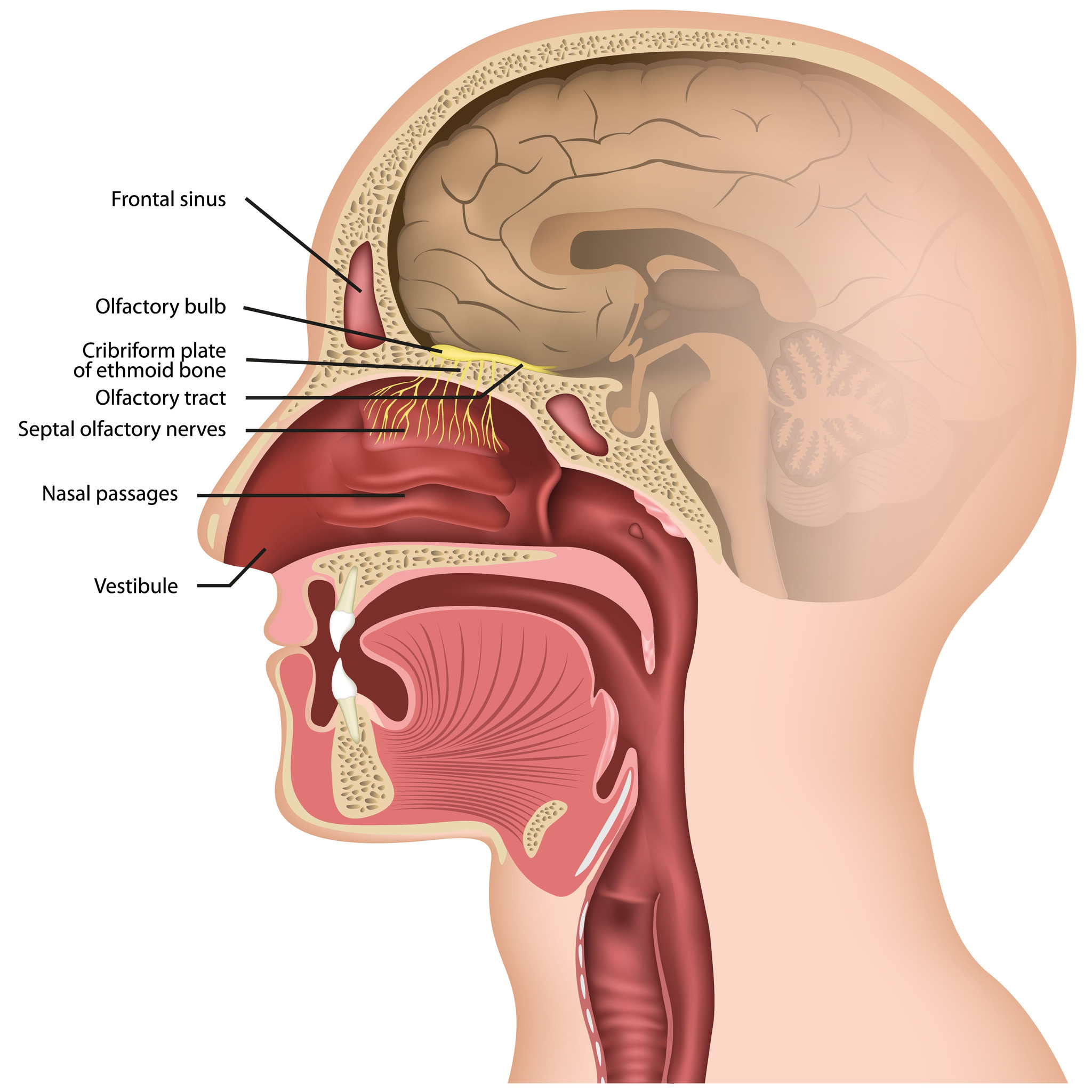
In rhinologic procedures, amniotic membrane has been explored for mucosal defects following functional endoscopic sinus surgery, septal surgery, turbinate surgery, and resection of nasal tumors. When placed over exposed cartilage, bone, or mucosal wounds, it may support epithelial migration and regeneration, maintain a moist wound environment, and reduce crust formation. In nasal septal perforation repair, it may be combined with a local mucosal flap to provide ECM support between tissue layers. Following sinus surgery, it may also help separate adjacent wound surfaces, maintain sinus ostial patency, and support restoration of mucosal integrity.
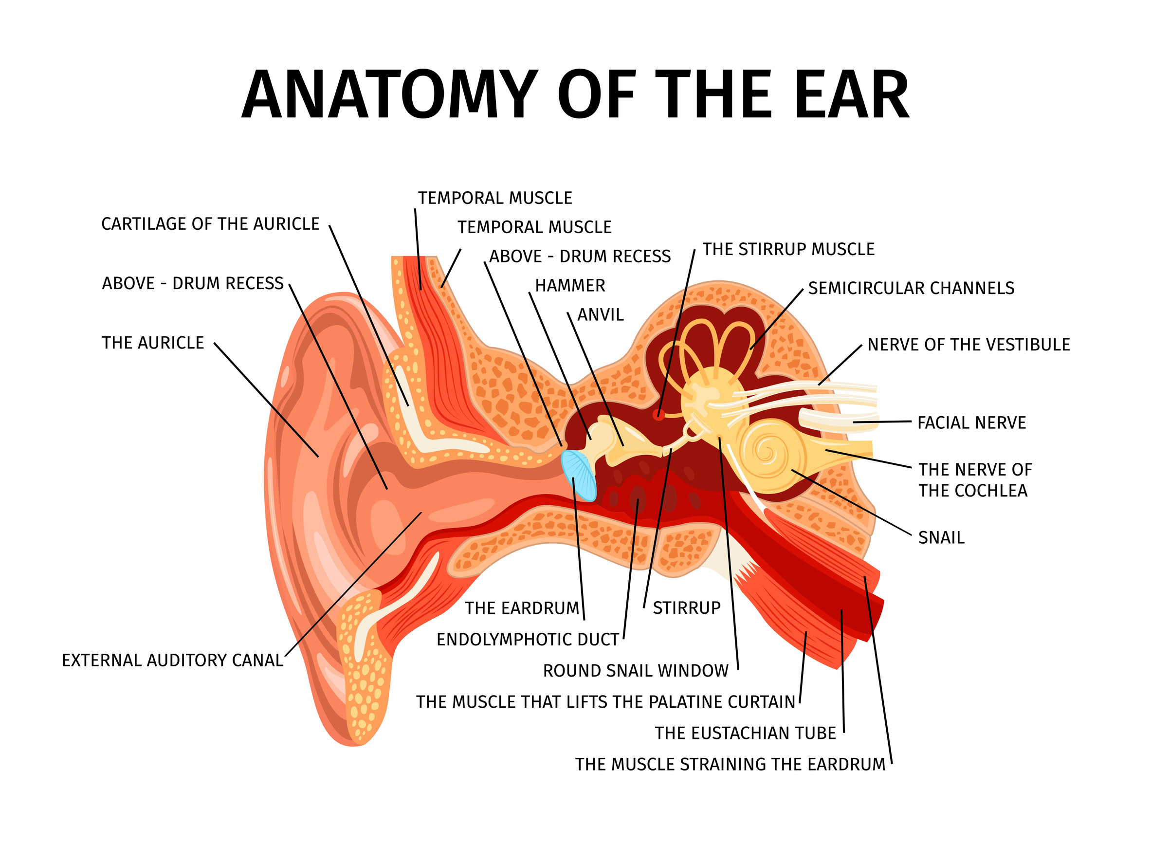
In otologic applications, amniotic membrane has been investigated for tympanic membrane perforation repair and tympanoplasty. Its thin and flexible structure allows it to conform to the tympanic annulus and perforation margins, where it may serve as a scaffold for keratinocyte and fibroblast migration and support reconstruction of the epithelial and fibrous layers. For external auditory canal defects, canaloplasty, or exposed bone following cholesteatoma removal, it may be placed over the wound to support epithelialization and reduce the risk of canal adhesion and stenosis.
In oral, pharyngeal, and head-and-neck surgery, amniotic membrane has been explored for oral mucosal defects, tonsillar surgical wounds, oropharyngeal tumor resection sites, and reconstruction of the soft palate or pharyngeal wall. Its conformability allows it to adapt to dynamic surfaces involved in swallowing and voice production while protecting injured tissue and supporting mucosal regeneration. For injuries involving the vocal folds, larynx, or tracheal mucosa, it may also serve as a wound covering and tissue-separation barrier to reduce adhesion between injured surfaces, preserve luminal structure, and support mucosal repair and re-epithelialization.
Amniotic tissue may be suitable for various delicate and spatially constrained procedures in otolaryngology. Its clinical use should be evaluated according to the defect location and depth, infection status, fixation method, patient condition, approved indications, and applicable regulations. Products should meet the relevant requirements for human tissue banking, manufacturing quality, and sterility. With appropriate surgical planning and fixation, amniotic tissue may serve as an adjunctive biomaterial for tissue coverage, mucosal repair, and adhesion management in otolaryngology.

